







What is microtia?
Microtia is a congenital malformation of the auricle —the visible part of the ear—: during pregnancy the ear does not complete its development, and the baby is born with an ear that is smaller, misshapen or, in the most severe form, only a remnant of skin and cartilage where the ear should be. It affects one ear more often than both, the right more often than the left, and is somewhat more common in boys.
It is not caused by anything the mother did or did not do. It can occur on its own or together with other developmental differences of the face, such as a less developed jaw or cheek on the same side. It is often associated with aural atresia —the ear canal is not open— which is why hearing is checked from the very first visit.
The grades of microtia
The shape of the ear weighs most in choosing a technique, so microtia is described by grade. The most widely used classification has four:
- Grade I: the ear is smaller than normal but keeps almost all its recognisable structures —the rim (helix), the inner folds, the concha, the lobe.
- Grade II: some structures are missing or deformed; often the upper third of the ear is folded or rolled over on itself and the ear looks short. Many severe «cup» or «constricted» ears fall here.
- Grade III: the classic form. A vertical, peanut-shaped remnant of skin and cartilage remains, almost always with a displaced lobe; the ear canal is usually closed.
- Anotia: complete absence of the ear. The least common form.
How an ear is reconstructed: the techniques
There is no single microtia operation: there is a spectrum, and what defines the plan is how much usable tissue the patient's ear has.
When almost the whole ear is missing (grade III and anotia), the standard is to build a rib-cartilage framework: cartilage is taken from the patient's own ribs, carved by hand into the shape of an ear —rim, folds, concha— and placed under the skin of the area. It is a staged operation: the framework goes in at a first stage; at a second, months later, the ear is lifted away from the head to give it projection and the groove behind it is created. The patient's own cartilage is living tissue: it grows with the child and is not rejected.
When the ear keeps a good part of its structures (grades I and II, constricted ears), it is often unnecessary to build a new ear; instead the existing ear is reconstructed: the rolled-over upper part is unfurled, the cartilage is redistributed and reinforced —sometimes with a cartilage graft from the ear itself or from the rib—, the rim is redefined and the skin is repositioned. Depending on the case, this can be done in a single operation.
Synthetic porous-polyethylene frameworks and skin-adhered silicone prostheses also exist; they are alternatives with specific indications, discussed at the consultation together with their advantages and risks.
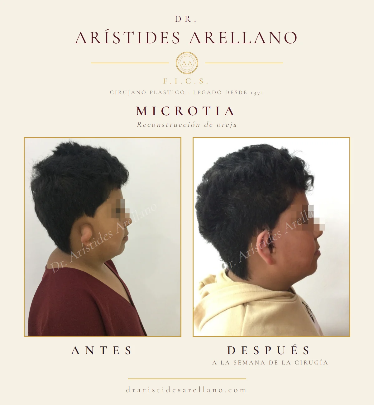
A real case: before and one week after
The case shown on this page is a school-age boy with microtia of the right ear. Before surgery the ear was short and its upper third was folded over on itself, with no defined rim and no inner folds; the lobe was preserved. The later photograph was taken one week after surgery, with the stitches still in place in front of the ear: the ear already has more height, a defined rim, a groove beneath the rim and a recognisable concha.
One week is very early to judge a result: swelling still changes the contour, and the shape continues to settle over the following months. Out of respect for his privacy, his eyes are hidden in the photographs. They are published with his family's consent. Results may vary from one person to another.
All surgery carries risk and requires a prior medical assessment. Results may vary according to each patient, their anatomy and their individual healing process.
At what age is microtia operated on?
Rib-cartilage reconstruction is usually planned from around age six, and many surgeons prefer to wait until eight to ten: before then there is not enough rib cartilage to carve a framework the size of an adult ear. By that age the healthy ear has also nearly reached its final size, which allows the two to be matched, and the child is entering the school years in which teasing starts to matter.
Ears that keep more structure can sometimes be corrected earlier, and in newborns with deformed ears —not true microtia— moulding devices take advantage of the first weeks of life. There is no upper age limit: reconstruction is also done in adults who were not operated on as children, although the cartilage is stiffer and is handled differently.
Hearing comes first
Before thinking about the shape of the ear, the child's hearing is checked. When there is canal atresia, the affected ear usually has a conductive hearing loss; the healthy ear usually hears normally. That assessment —audiometry, evoked potentials and, when indicated, a CT scan of the ear— is performed by an ENT specialist or audiologist, to whom the clinic refers the patient.
The order matters: if opening the canal or fitting a hearing device is planned later, the skin and cartilage of the reconstruction are planned together with the ear specialist, so that no options are closed off.
How the case is assessed at the clinic
At the consultation Dr. Arístides Arellano examines the affected ear and the healthy one, measures the difference in height and position between them, checks the quality of the skin in the area —scars, hair, thickness— and the development of the face on the same side, and reviews the hearing tests. From that the grade, the technique and the number of surgical stages are defined, and the family is told what they can and cannot expect.
When the patient is a minor, every decision is made with the parents or guardians, and the child takes part in the conversation according to their age: ear reconstruction works best when the patient wants it.
Cost of ear reconstruction
We do not publish a single figure for microtia because there is no standard case: the cost depends on the technique, on whether rib cartilage is needed, on the number of surgical stages, the type of anaesthesia and the hospital and anaesthetist fees. The definitive cost is set at a private assessment consultation with Dr. Arístides Arellano —$800 MXN, charged separately. For minor deformities of the auricle, such as prominent ears, see otoplasty.
Microtia, otoplasty and reconstruction: how they relate
They are three different things and are best not confused. Otoplasty corrects ears that are well formed but prominent; microtia is an ear that did not form completely. Eyelid, ear and nose reconstruction covers surgery of these three facial units when they are lost to skin cancer or an accident, and congenital defect correction groups the birth malformations treated by plastic surgery, of which microtia is one of the best known.
Who performs the surgery
Dr. Arístides Arellano Huacuja is a plastic, aesthetic and reconstructive surgeon in Puebla. He qualified as a physician and surgeon at Universidad Autónoma Metropolitana Xochimilco (1986) and practises under Mexican professional licence D.G.P. 1125959 and specialty licence 0002008; he trained in his specialty at Hospital General de Agudos Dr. Abel Zubizarreta, in Buenos Aires, between 1987 and 1989. He has been a Fellow of the International College of Surgeons (F.I.C.S.) since 1994 and holds US patent 5,928,158 (USPTO, 1999).
He practises at the Clínica Dermatológica y Cirugía Estética de Puebla, founded in 1971 by his father, the dermatologist Dr. Francisco Arellano Ocampo; he directs it today, in the second generation. The clinic is at Calle 20 Sur 2539, Col. Bellavista, 72500 Puebla, Pue.; hours are Monday to Friday 8:00–20:00 and Saturday 8:00–14:00, and assessments are booked on +52 221 155 2228.
Trust is verified, not promised. All 210 scanned documents —degree, licences, diplomas, publications and certificates, from 1977 to 2025— are open in Dr. Arístides Arellano’s credential archive, alongside an explanation of the difference between a specialty licence and a board certification.
Recovery · step by step
- First daysProtective dressing over the ear and, if rib cartilage was taken, chest discomfort that is usually the most bothersome part. Prescribed pain relief and relative rest at home.
- Week 1Check-up and dressing change at the clinic. The ear looks swollen and the stitches are still in; the shape is not yet final.
- Weeks 2 to 3Stitches removed according to progress. Return to school or work, avoiding knocks and pressure on the ear while sleeping.
- Weeks 4 to 6Physical activity resumes; contact sports wait until the surgeon allows them.
- 3 to 6 monthsSwelling settles and the relief of the ear becomes defined. If the plan is staged, the next operation is scheduled once the tissues have matured, usually several months later.
Risks and contraindications
Ear reconstruction is delicate surgery and, like any operation, carries specific risks that are reviewed with the family before deciding:
- Infection: uncommon, but in a reconstruction it can threaten the cartilage; it is watched closely and treated immediately.
- Skin compromise or loss: the skin covering the cartilage is thin; if its circulation suffers, the framework can become exposed and need a coverage operation.
- Haematoma: a collection of blood under the skin that may need draining.
- Donor-site discomfort: when rib cartilage is used, chest pain in the first weeks and a scar on the chest; rarely, an injury to the pleura that is identified and resolved during surgery.
- Cartilage resorption or loss of definition over time, which can make the relief less visible.
- Differences in size, position or projection compared with the healthy ear; a reconstructed ear is not identical to a natural one.
- Scars: around the ear and, where applicable, on the chest; their quality also depends on each patient's skin.
- Anaesthetic risks: those of any procedure under general anaesthesia; assessed with the medical history and pre-operative tests.
Surgery is postponed or reconsidered in any of these situations:
- An age or stage of development that does not yet provide enough cartilage for the planned technique.
- Active infection of the ear or the surrounding skin, or a significant respiratory illness around the date of surgery.
- Uncontrolled illness or clotting disorders.
- A pending hearing assessment when canal surgery is planned: the ear and the hearing are planned together.
- A patient old enough to have a view who does not yet want the operation.
Frequently asked questions
What is microtia?
It is a congenital malformation in which the ear does not finish forming during pregnancy: the baby is born with an ear that is smaller, has incomplete folds or is only a remnant of skin and cartilage. It usually affects one ear and is often accompanied by a closed ear canal, so hearing is always checked.
At what age is microtia operated on?
Rib-cartilage reconstruction is usually considered from around age six, when there is enough cartilage to carve the framework; many surgeons prefer eight to ten. Ears that keep more structure can sometimes be corrected earlier, and there is no upper age limit for adults. The exact timing is set at the consultation.
How is an ear with microtia reconstructed?
It depends on the grade. When almost the whole ear is missing, a framework is carved from the patient's own rib cartilage and placed under the skin over several stages. When the ear keeps a good part of its structures, the existing ear is often reconstructed: the rolled part is unfurled, the cartilage reinforced and the rim redefined, sometimes in a single operation.
Does microtia surgery improve hearing?
Reconstructing the auricle improves the shape of the ear, not hearing. If there is hearing loss from a closed canal, it is assessed separately by an ENT specialist or audiologist, who may propose opening the canal or a hearing device; when planned, it is coordinated with the reconstruction.
How many operations are needed?
With rib cartilage, usually two or more stages several months apart. Ears with more structure can be corrected in a single operation, sometimes with a later touch-up. The exact number of stages is set at the consultation, according to the grade and the skin of the area.
Does the reconstructed ear grow with the child?
When it is built from the patient's own cartilage, yes: it is living tissue that keeps pace with growth, which is why surgery is timed for when the healthy ear has nearly reached adult size. A synthetic framework does not grow.
How much does microtia ear reconstruction cost in Puebla?
There is no single figure: it depends on the technique, whether rib cartilage is used, the number of stages and the hospital and anaesthesia fees. The definitive cost is set at a private assessment consultation with Dr. Arístides Arellano, $800 MXN, charged separately.
Before and after
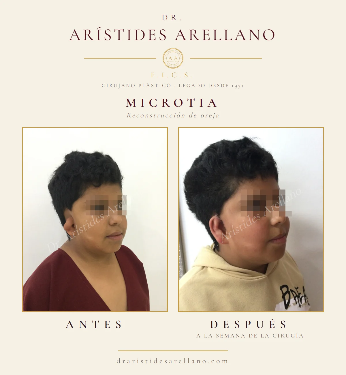
Right-ear microtia, three-quarter view. Before and one week after surgery, stitches still in place; the shape keeps settling over the following months. The eyes are hidden to protect the patient’s privacy.
Photographs of real patients, published with their written informed consent. Results may vary from person to person; these images are not a promise of outcome.
Every case is different. Discuss yours in a personal consultation.
Book a consultation on WhatsAppEvery figure published on this site is approximate and is not a quote. Any procedure or treatment first requires a private assessment consultation with Dr. Arístides Arellano —$800 MXN, charged separately— and it is at that consultation that the definitive cost of your case is determined.
Educational content; it does not replace a medical consultation. Results may vary from person to person.